
Fig. 2.
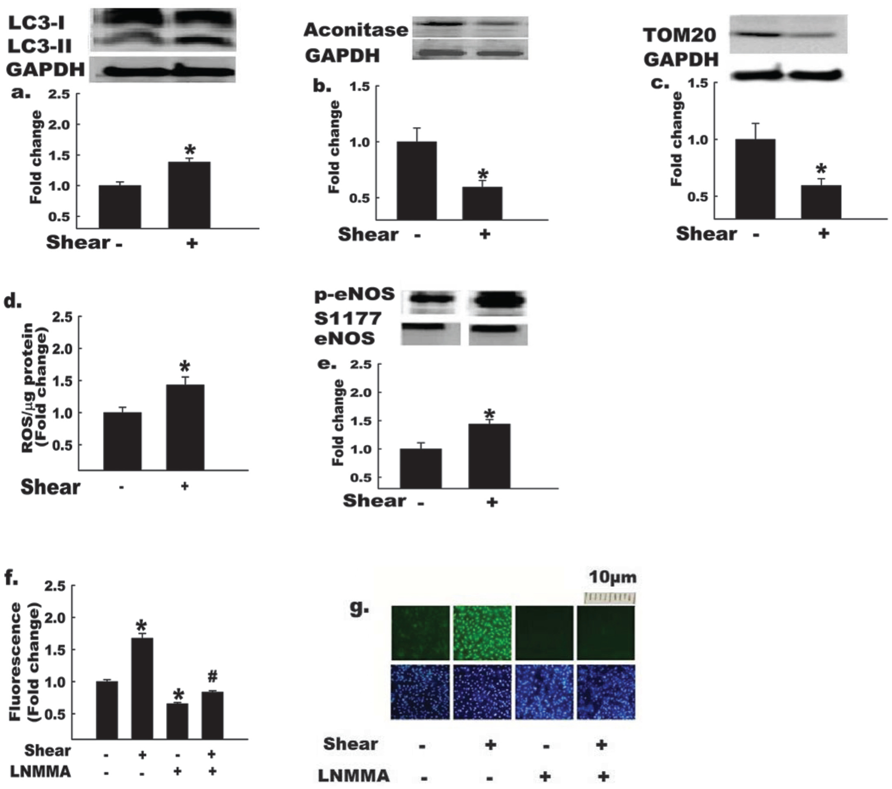
Autophagy and nitric oxide (NO) bioavailability are elevated in endothelial cells exposed to shear stress. Bovine aortic endothelial cells (BAECs) were exposed to shear stress (shear +) or static conditions (shear −) for 3 h, as indicated. GAPDH, glyceraldehyde-3-phosphate dehydrogenase. Shear + BAECs show increased (*, p < 0.05) LC3-II:LC3-I ratio (a), and decreased protein levels of mitochondrial markers, m-aconitase (b), and TOM20 (c); in addition, shear + BAECs exhibit increased reactive oxygen species (ROS) production (d), p-eNOS S1177 : total eNOS (e), and NO production (mean data (panel f), representative images of DAF-FM fluorescence (top row, panel g)) in comparison with shear – BAECs (n = 4–6 per experiment). Treatment of BAECs with the NO synthase inhibitor, monomethyl-L-arginine (L-NMMA), reduced (p < 0.05) NO production ± shear stress (mean data (panel f), representative images of DAF-FM (panel g, top row) and DAPI nuclear fluorescence (panel g, bottom row)). The bottom row of panel g indicates that cell density was similar among treatments. For Figs. 2a, 2b, 2c, and 2e, the histograms (below) represent the mean ± SE of densitometry (above). *, p < 0.05 for shear effect; #, p < 0.05 for L-NMMA effect. For Figs. 2a, 2b, 2c, and 2e, each n refers to one 10 cm petri dish. For Figs. 2d, 2f, and 2g, each n refers to one well of a 6-well plate.