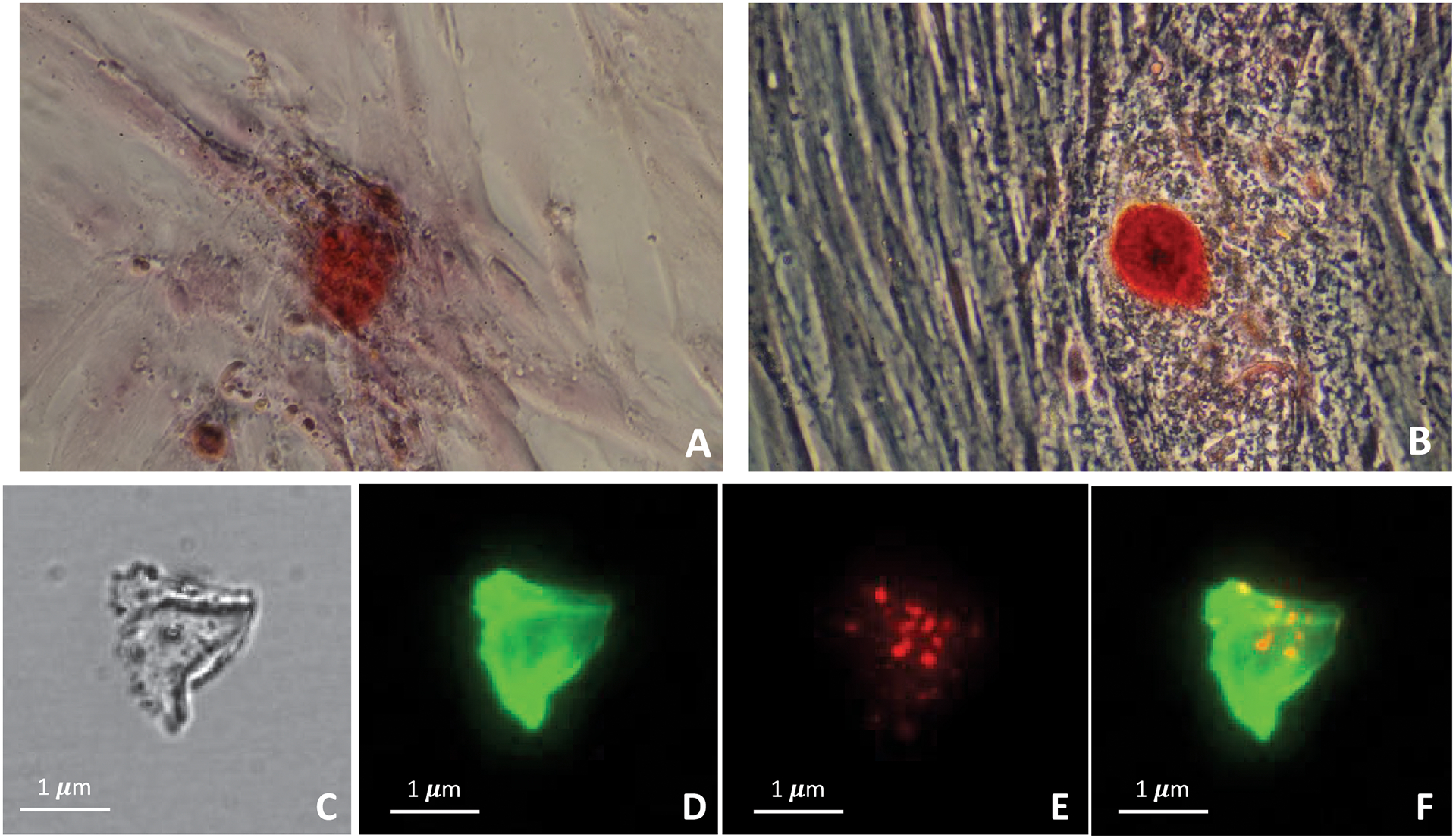
Figure 5.

Alizarin red staining of VSMCs differentiated in chondrocyte/osteoblast phenotype under different conditions for 14 days: (A) 10 μg/mL ascorbic acid and 5 mM Na2PO4 and (B) 50 μg/mL ascorbic acid and 5 mM Na2PO4 (magnification factor 200x). Colocalization analysis by immunofluorescent staining between proteoliposomes and type II collagen matrix produced from culture of osteo/chondrocytic cells: (C) white light image of native collagen matrix; (D) type II collagen detected by anti-collagen type II antibody (green fluorescence); (E) Bound AnxA5 9:1 DPPC:DPPS proteoliposomes (1.87 μg of protein content) labeled with Rhodamine to type II collagen matrix (red fluorescence); (F) Co-localization of bound proteoliposomes with type II collagen (yellow).
