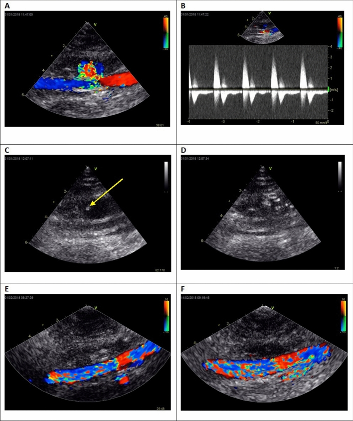
Figure 2.
The example of the UGTGI procedure. (A) Ultrasound two-dimensional longitudinal image with color Doppler before psA embolization. (B) Pulse-wave Doppler image of inflow and outflow from the sac of psA, LEVI = 0.18. (C) Inserting a needle into the psA sac. The arrow indicates the tip of the needle. (D) Injection of TG into the psA sac. (E) Ultrasound image after the embolization of psA. (F) Follow-up ultrasound examination after 2 weeks. psA—pseudoaneurysm, UGTGI—ultrasound-guided tissue glue injection, LEVI—late to early velocity index, TG—tissue glue.

