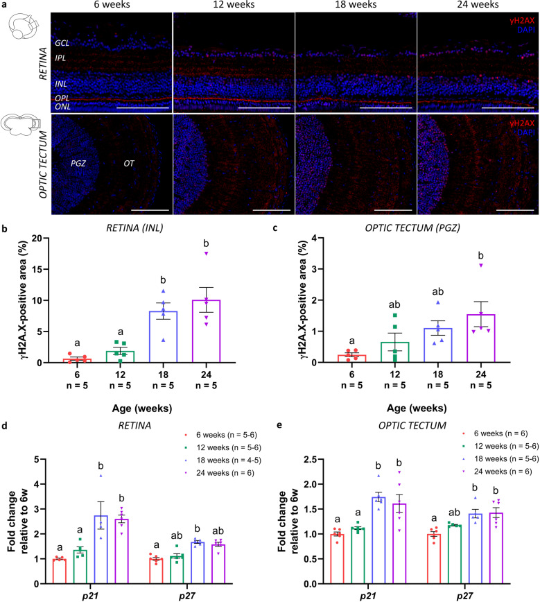
Fig. 3. Accumulation of DNA damage and elevated levels of cell cycle regulators in the senescent killifish visual system.
a Microscopic images of γH2AX-labeled retinal and brain cryosections show a clear accumulation of DNA damage in the nuclei, particularly visible in the GCL and INL of the retina, and the PGZ of the optic tectum. Scale bar = 100 µm. b, c Quantification of the γH2AX-immunopositive area within a predefined region of the retinal INL and tectal PGZ reveals a significant age-related increase. Values represent mean ± standard error of mean, n = 5. d, e Real-time quantitative PCR for cyclin-dependent kinase inhibitors p21 and p27, both regulators of cell cycle progression and typical markers for cellular senescence, reveals a significant rise in gene expression levels with increasing age, both in the retina (d) and optic tectum (e). Values are mean fold change values relative to values of 6-week-old fish ± standard error of mean, n ≥ 4. Statistical significance between different conditions is shown using different letters. γH2AX gamma H2A histone family member X, DAPI 4′,6-diamidino-2-phenylindole, GCL ganglion cell layer, IPL inner plexiform layer, INL inner nuclear layer, OPL outer plexiform layer, ONL outer nuclear layer, PGZ periventricular gray zone, OT optic tectum.

