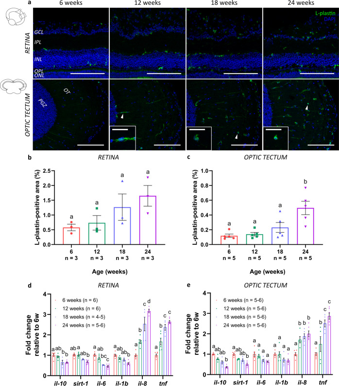
Fig. 6. Inflammaging in the senescent killifish visual system.
a Representative images of the retina and the optic tectum immunolabeled for the pan-leukocyte marker L-plastin illustrate a visible increase in microglial/macrophage number with increasing age. Moreover, an age-dependent change in morphology is observed in 18-week- and 24-week-old fish, in which microglia seem to transform from a ramified-like toward ameboid-like form (white arrowheads in optic tectum; magnified in boxes; scale bar = 20 µm). Scale bar = 100 µm. b, c Quantification of the area occupied by immunostained leukocytes over the total analyzed area demonstrates an increase in the microglial/macrophage area fraction with increasing age. Values are shown as mean ± standard error of mean, n ≥ 3. d, e Quantification of mRNA levels of inflammatory molecules, such as il-10, sirt-1, il-6, il-1b, sirt-1, il-8, and tnf, in both retinal and tectal lysates reveals significant changes in the expression profile in the retina as well as optic tectum with advancing age. While the expression values of il-10, sirt-1, il-6, and il-1b decline upon aging, levels of il-8 and tnf increase in older fish. Values represent mean fold change values relative to values of 6-week-old fish ± standard error of mean, n ≥ 4. Statistical significance between different conditions is indicated using different letters. DAPI 4′,6-diamidino-2-phenylindole, GCL ganglion cell layer, NFL nerve fiber layer, IPL inner plexiform layer, INL inner nuclear layer, OPL outer plexiform layer, ONL outer nuclear layer, PRL photoreceptor segment layer, SO stratum opticum, SFGS stratum fibrosum et griseum superficiale, SGC stratum griseum centrale, PGZ periventricular gray zone, OT optic tectum, il-10 interleukin 10, il-1b interleukin 1β, il-6 interleukin 6, sirt-1 sirtuin 1, il-8 interleukin 8, tnf tumor necrosis factor.

