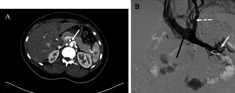
Figure 2.
Left renal vein compression associated with symptoms of left flank pain and hematuria. (A) Computed tomography (CT) demonstrates compression of the left renal vein (white arrow) over the abdominal aorta. (B) Venography demonstrates contrast attenuation over the abdominal aorta (black arrow), renal hilar varices (white arrow), and ascending collaterals (dashed white arrow) consistent with renal vein compression. The Symptoms-Varices-Pathophysiology (SVP) classification is S1V1PLRV,O,NT.

