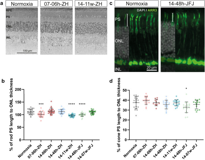
Figure 5.
Photoreceptor segment lengths in hypoxia. (a) Representative retinal morphologies and (b) graph showing the ratio (%) of rod photoreceptor segment length to outer nuclear layer (ONL) thickness for indicated hypoxic conditions. (c) Representative immunostaining for cone arrestin (ARR3, green) and (d) the percentage of cone photoreceptor segment length to outer nuclear layer (ONL) thickness. Cell nuclei were counterstained with DAPI (grey). One-way ANOVA with Dunnett’s multiple comparison test was used to compare each condition to control (normoxia) *p < 0.05; ***p < 0.001; ****p < 0.0001. RPE: retinal pigment epithelium; PS: photoreceptor segments; ONL: outer nuclear layer; INL: inner nuclear layer; GCL: ganglion cell layer. Scale bars as indicated.

