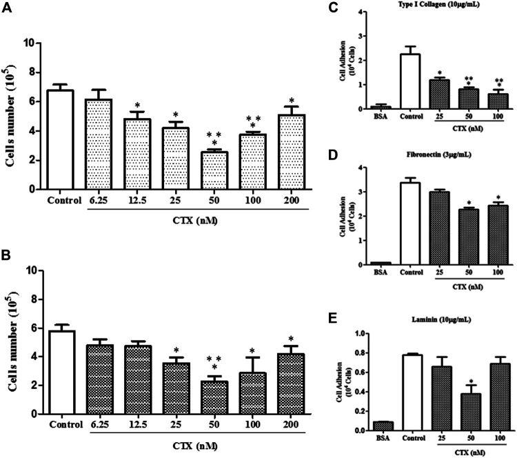
FIGURE 1.
Effect of CTX on t.End.1 cell proliferation and adhesion to extracellular matrix ligands. t.End.1 cells (5 × 10⁴ cells/well) were incubated in the presence of different CTX concentrations (6.25, 12.5, 25, 50, 100 and 200 nM) for 24 h (A) or were previously incubated with the different concentrations for only 1 h and then washed and incubated for another 24 h only in fresh culture medium (B). Cell proliferation was assessed after 24 h by cell counting. The data are presented from three distinct experiments run in octoplicate and are expressed as mean ± s.e.m. *p < 0.05 compared to control group, **p < 0.05 compared to other CTX concentrations. For adhesion assay, t.End.1 cells pretreated for 1 h with CTX (25, 50 and 100 nM) were washed and added (100 µl) to Maxsorp plates (Nunc®) containing 96 wells, previously sensitized with the different ligands of matrix: type I collagen (10 µg/ml) (C); fibronectin (3 µg/ml) (D) and laminin (10 µg/ml) (E). After 1 h, adhered cells were evaluated by MTT assay. The values obtained were fed into GraphPad INSTAT program V2.01 for conversion of optical density (OD) in the number of adhered cells. The data are presented from three distinct experiments run in sextuplicate and are expressed as mean ± s.e.m. *p < 0.05, compared to control group and **p < 0.01, significantly different from mean values for groups to their respective CTX-treated cells.

