Figure 2.
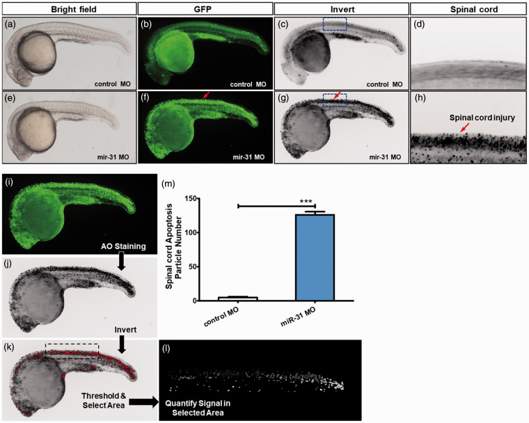
miR-31 knockdown induces CNS-specific apoptosis. Embryos injected with control or miR-31 MO were stained with acridine orange (AO) at 26 hpf. The apoptotic cells are shown as bright green spots or black spots. Less bright homogenous green or black is unspecific background staining. (a–d) Controls exhibited few or no apoptotic cells in the central nervous system (CNS). In contrast, significantly increased staining was observed throughout the CNS in zebrafish injected with miR-31 MO (f–h, red arrows). The blue box area is shown with higher magnification in the right panels. (i–m) Quantification of apoptosis number in the spinal cord shows a 25-fold increase in miR-31 morphants (n = 10) at 26 hpf. A-H: lateral view, anterior, left; hpf: hours post-fertilization. Columns, mean; bars, SEM. (A color version of this figure is available in the online journal.)

