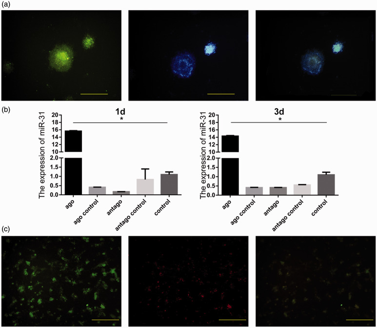
Figure 4.
Identification of neural stem cells cultured in vitro and detection of miR-31 after transfection. Green cells are positive for Nestin; blue indicates cell nuclei stained with Hoechst. They are overlapping after merging (a). miR-31 was overexpressed in the agomir group and lowly expressed in the antagomir group (b). Positive cells for miR-31 are red, and they overlap with Nestin-positive cells (c). Scale bar = 10 µm. *P < 0.05. Columns, mean; bars, SEM. (A color version of this figure is available in the online journal.)

