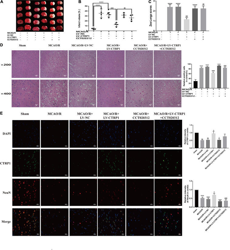
FIGURE 6.
The protective effect of CTRP1 in CIRI was abolished by CCT020312. (A,B) TTC staining and infarct volume in the brain. n = 4 per group. (C) Zea-longe scores. n = 13 per group. (D) HE staining and relative positive cells in the cortex. n = 3 per group. The representative images were acquired under × 200 magnification, scale bars = 100 μm, × 400 magnification, scale bars = 50 μm. (E) Double labeling immunofluorescence staining in the cortex (× 400 magnification). n = 3 per group. The representative images were acquired under × 400 magnification, scale bars = 50 μm. ****p < 0.0001, ∗∗p < 0.01, *p < 0.05 vs. sham group, ####p < 0.0001, ###p < 0.001, ##p < 0.01, #p < 0.05 vs. MCAO/R + LV-NC group; @@p < 0.01, @p < 0.05 vs. MCAO/R + LV-CTRP1 group.

