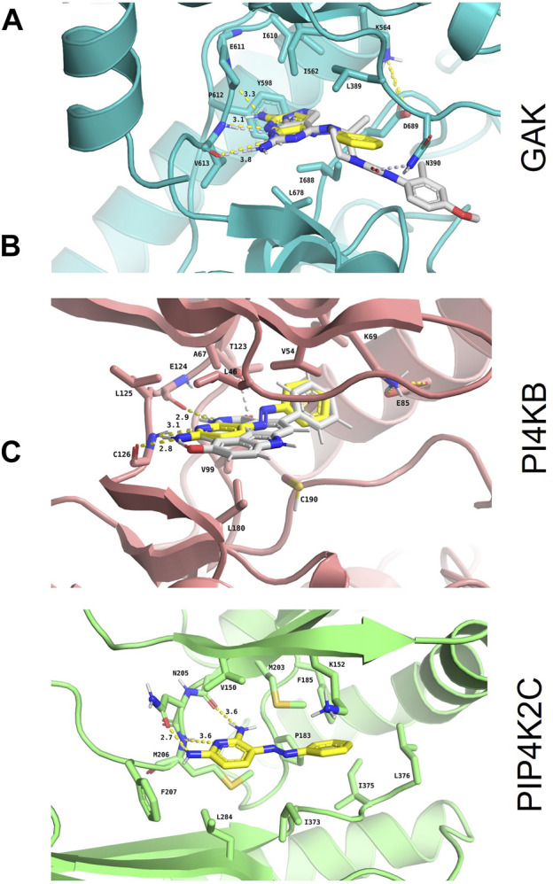
FIGURE 3.

In silico confirmation of phenazopyridine binding to the three selected kinases. Binding mode and interaction diagrams of phenazopyridine within (A) human cyclin-G-associated kinase (GAK; PDB identifier: 4Y8D), (B) phosphatidylinositol 4-kinase β (PI4KB; PDB identifier: 6GL3), and (C) phosphatidylinositol-5-phosphate four kinase type 2 gamma (PIP4K2C; PDB identifier: 2GK9). The atoms of phenazopyridine are shown as sticks with yellow carbons and the interacting residues of each protein as sticks with orange (A), blue (B), and brown carbons (C), respectively. For the holo crystal structures (A) and (B), the carbon atoms of the respective co-crystallized ligands are displayed with white sticks. H-bonds are shown as dashed yellow lines, and their lengths are indicated in Ångströms. Binding site residues directly interacting with phenazopyridine and the catalytic Lys are labelled as well.
