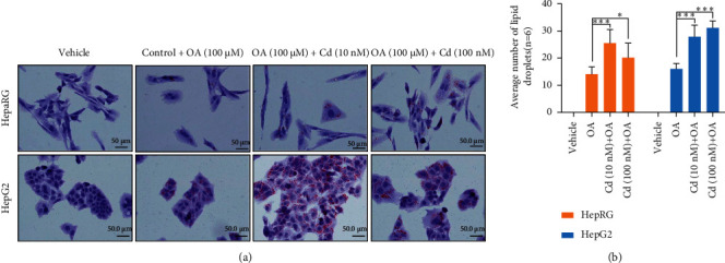
Figure 3.

Cd increases HCC cell steatosis. (a) Normal liver HepaRG cells and HCC HepG2 cells were exposed with 10 nM and 100 nM of Cd for 30 h and cells were treated with oleic acid (100 μM) for an additional 24 h. Cells were fixed with paraformaldehyde and stained with oil red O and images were captured and presented. (b) The effect of Cd on oleic acid-mediated lipid droplet formation was quantified and plotted. ∗P < 0.05, ∗∗∗P < 0.001 compared with oleic acid- (OA-) treated cells.
