
Figure 1.
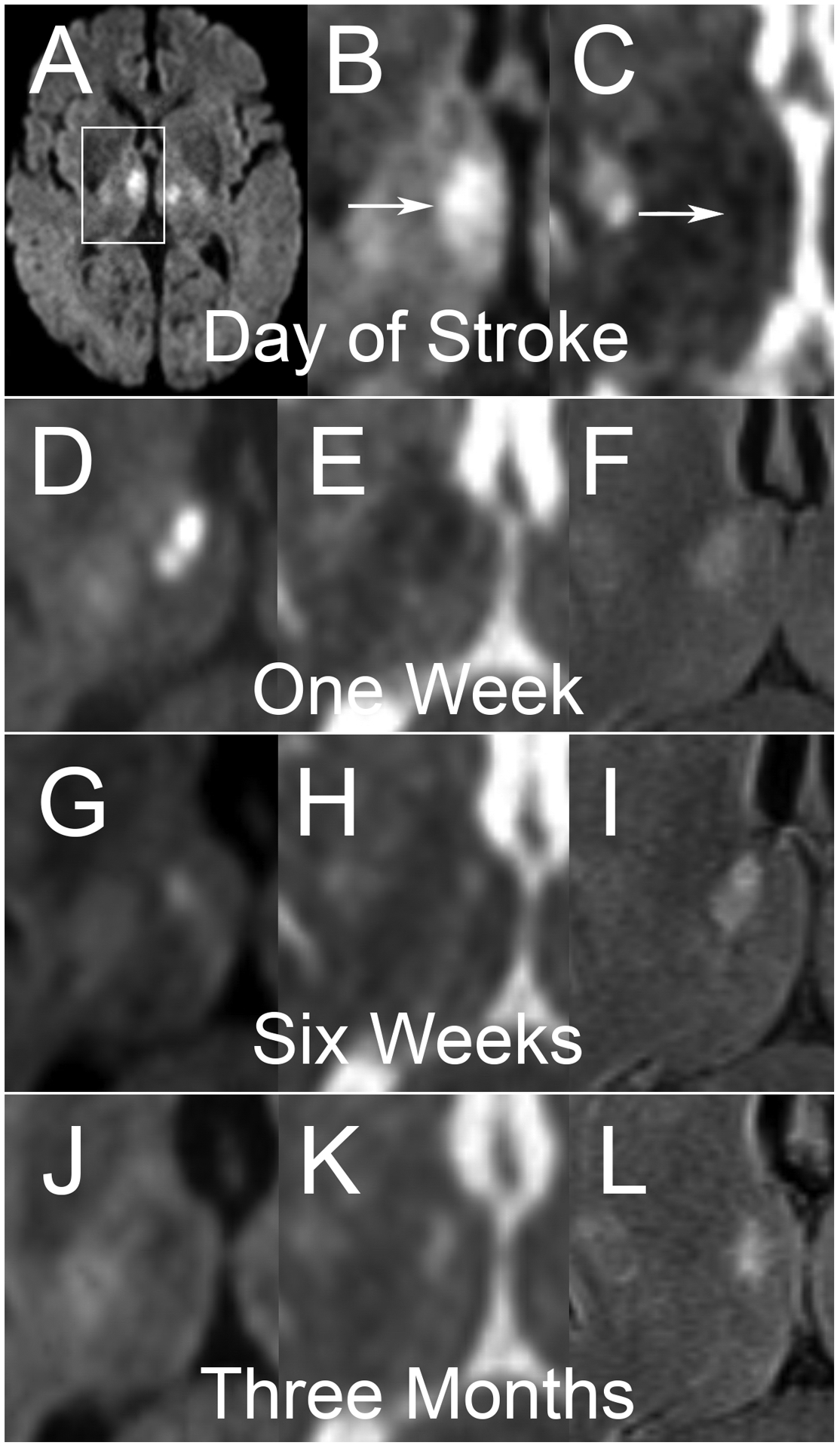
A middle-aged man presented with altered mental status and was found to have bilateral thalamic infarcts on DWI (A). White box demonstrates region of interest for subsequent cropped axial images of the right deep gray structures. Sequential studies with DWI (B, D, G, J), ADC (C, E, H, K) and FLAIR (F, I, L) sequences are shown. Imaging on the day of the infarct (A, B, C) demonstrates hyperintense DWI signal with hypointense ADC signal. Of note, FLIAR imaging was not performed at this time. At one week, DWI (D) demonstrates more pronounced hyperintensity with clear corresponding hypointensity on ADC (E) and hyperintensity on FLAIR. At six weeks, very little DWI (G) hyperintensity persists, which matches corresponding ADC (H) that is now hyperintense, consistent with T2 shine-through confirmed on FLAIR (I). At three months, All DWI (J) hyperintensity has resolved, with some hypointensity seen in the area of the chronic infarct. ADC (K) and FLAIR (L) both demonstrate hyperintense signal.