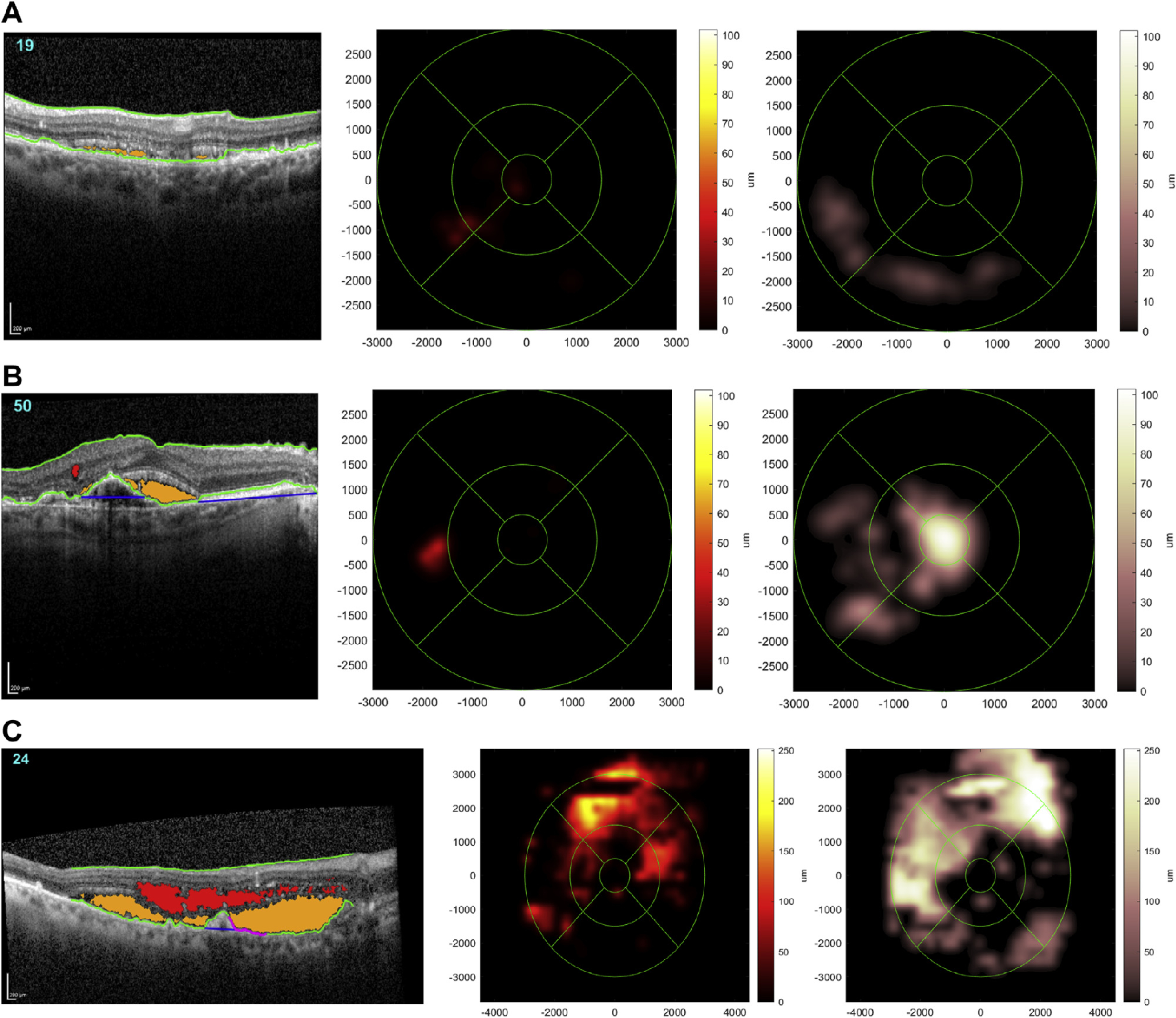
Figure 2.

Representative examples of spectral domain OCT (SD-OCT) scans where the Notal OCT Analyzer (NOA) correctly identified the presence of retinal fluid. A representative B-scan is shown (left) in each case, demonstrating how the NOA identifies and color-codes intraretinal fluid (red) and subretinal fluid (orange) on every B-scan. Also shown for each case are the 2 Early Treatment Diabetic Retinopathy Study grid heatmaps automatically generated by the NOA, 1 for intraretinal fluid (middle) and 1 for subretinal fluid (right); these provide rapid visualization of the location and extent of fluid separately for each tissue compartment. The total volumes of intraretinal fluid and subretinal fluid estimated by the NOA are displayed in nanoliters. A, Low total fluid volume (22 nl). B, Moderate total fluid volume (136 nl). C, High total fluid volume (3374 nl).
