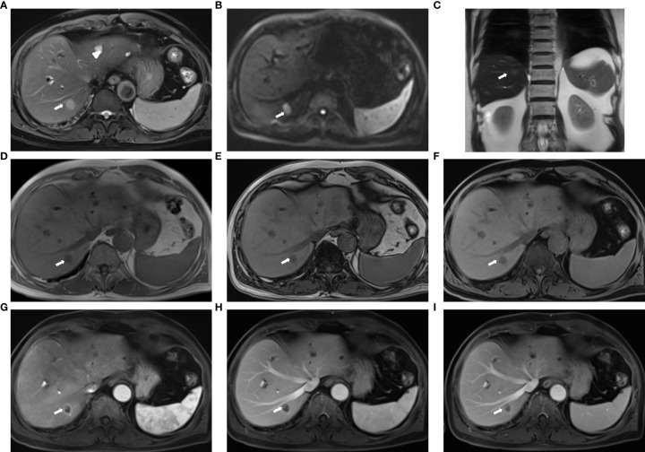
Figure 2.
Sample images acquired with the fast liver workflow of a 74-year-old man with liver metastasis (primary: colon cancer): (A) T2-weighted BLADE; (B) DWI, b = 1000 s/mm2; (C) T2-HASTE; (D, E) in-phase and out-of-phase DIXON VIBE; (F) water images of DIXON VIBE; (G–I) contrast-enhanced T1WI VIBE with arterial-phase (G), venous-phase (H), and delayed-phase (I). The metastasis (arrow) in liver segment VII appeared as slightly high signal intensity on the T2 BLADE and T2 haste image, whereas the hepatic cysts [arrowhead in (A)] in liver segments II and IV show high signal intensity in T2 BLADE. The metastasis is hyperintense in DWI and has low intensity in T1WI. Annular enhancement is visible in the enhanced MR imaging relative to the surrounding liver parenchyma.

