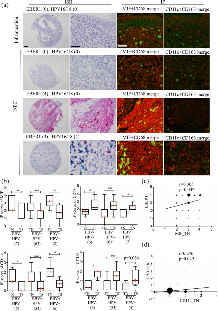
Fig. 2.
Correlation between virus infection status and macrophage migration inhibitory factor (MIF), macrophage markers. (a) In situ hybridization (ISH) images of EBER1 (red) and HPV16/18 (green), and immunofluorescence (IF) images of MIF (red) and macrophage markers, CD68 (green), CD11c (red), and CD163 (green). (b) Graphic presentation of the IF scores for MIF, CD68, CD11c, and CD163 in subgroups by EBV and HPV status. “TN” indicates tumor nest, “TS” indicates tumor stroma. #: p < 0.05, ##: p < 0.01, ##: p < 0.001, analyzed by paired test. The red cross in the graphic represents the median. (c) Correlation between EBER1 level and MIF score in tumor nest among 78 nasopharyngeal carcinoma (NPC) cases. (d) Correlation between HPV16/18 level and CD11c score in tumor nest among 65 NPC cases. The size of the dot represented the number of cases. For ISH low magnificent image, the scale bar represents 100 μm, and the other scale bar represents 40 μm

