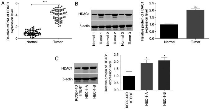
Figure 1.
Assessment of HDAC1 expression levels in endometrial cancer and matched-normal tissues. (A) Reverse transcription-quantitative PCR and (B) western blot analysis revealed that HDAC1 expression at both the mRNA (64 paired tumors) and protein (3 paired tissues) level was significantly increased in endometrial cancer tissues, compared with normal tissues. Normal 1/2/3 and Tumor 1/2/3 are representative of the normal tissue and cancer tissue samples from 64 cases. ***P<0.001, paired Student's t-test. (C) Protein levels of HDAC1 in HEC-1-A, HEC-1-B and KC02-44D hTERT cells were determined by western blotting. N=3; *P<0.05, ANOVA followed by Tukey test. HDAC1, histone deacetylase 1.

