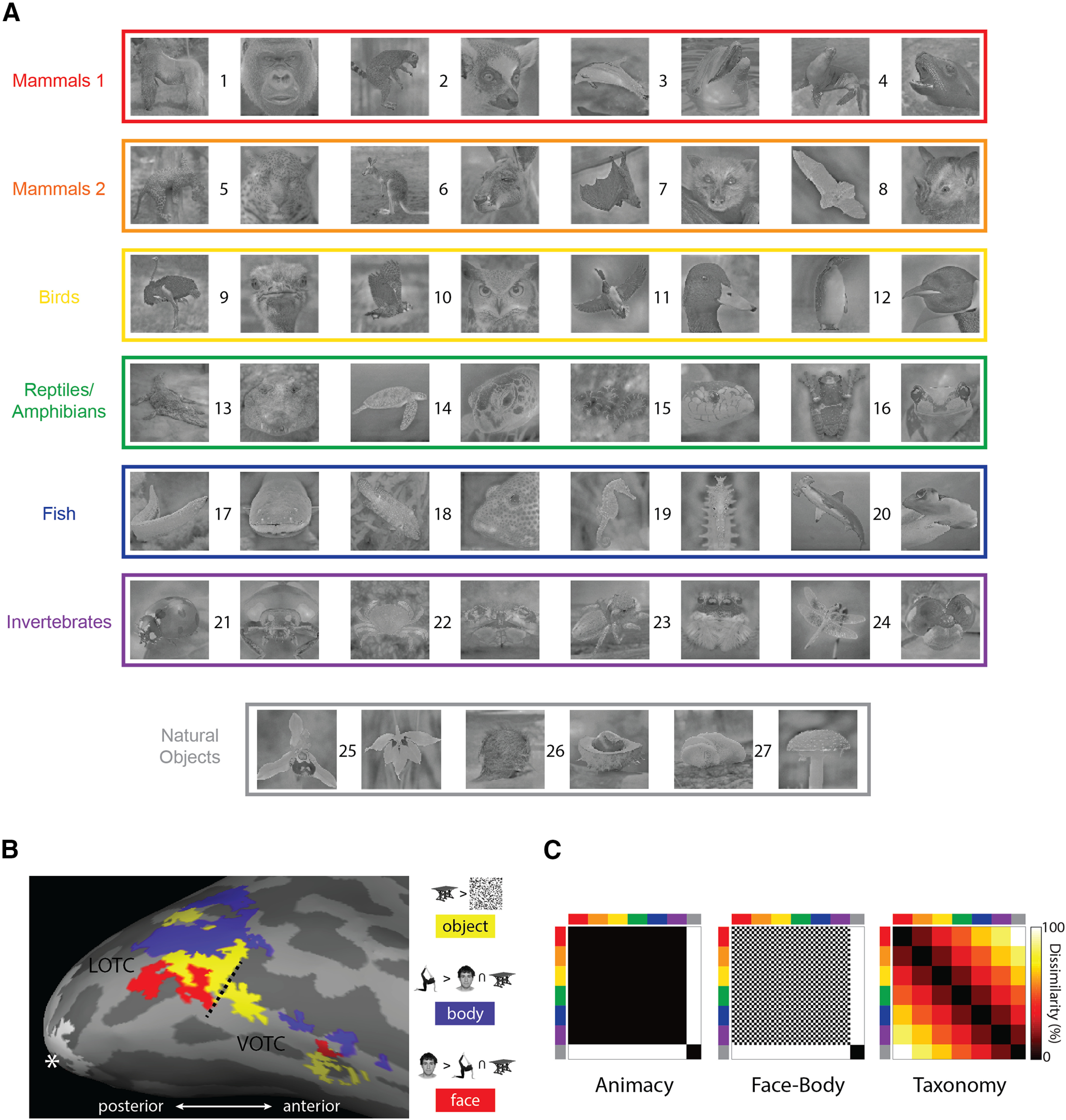
Figure 1.

Core features of the experimental design. A, All 54 natural image stimuli, color coded based on the taxonomic hierarchy. Numbers identify individual animals/natural object types: (1) gorilla, (2) lemur, (3) dolphin, (4) seal, (5) leopard, (6) kangaroo, (7) flying fox, (8) bat, (9) ostrich, (10) owl, (11) duck, (12) penguin, (13) crocodile, (14) turtle, (15) snake, (16) frog, (17) eel, (18) reef fish, (19) seahorse, (20) shark, (21) ladybug, (22) crab, (23) spider, (24) dragon fly, (25) orchid, (26) fruit, and (27) mushroom. B, The results of the functional contrasts used in the study to define the ROIs, for one representative participant, mapped onto the inflated cortex using FreeSurfer (Fischl, 2012). The hashed line indicates the boundary between the lateral and ventral masks defined using the Anatomical Toolbox (Eickhoff et al., 2005). The white asterisk indicates the occipital pole, and the white patch is the EVC. C, The three main model RDMs used throughout the study. The axes of the RDMs are color coded to reflect the taxonomic hierarchy for the stimuli.
