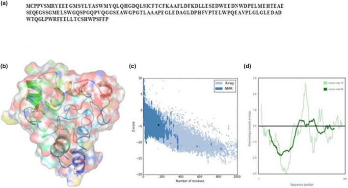
FIGURE 1.

3D structure of TEX19 protein. (a) The protein sequence of TEX19. (b) 3D structure. (c) Z‐scores of all protein chains in PDB, which are determined by X‐ray crystallography (light blue) or NMR spectroscopy (dark blue). (d) Validation of the developed model
