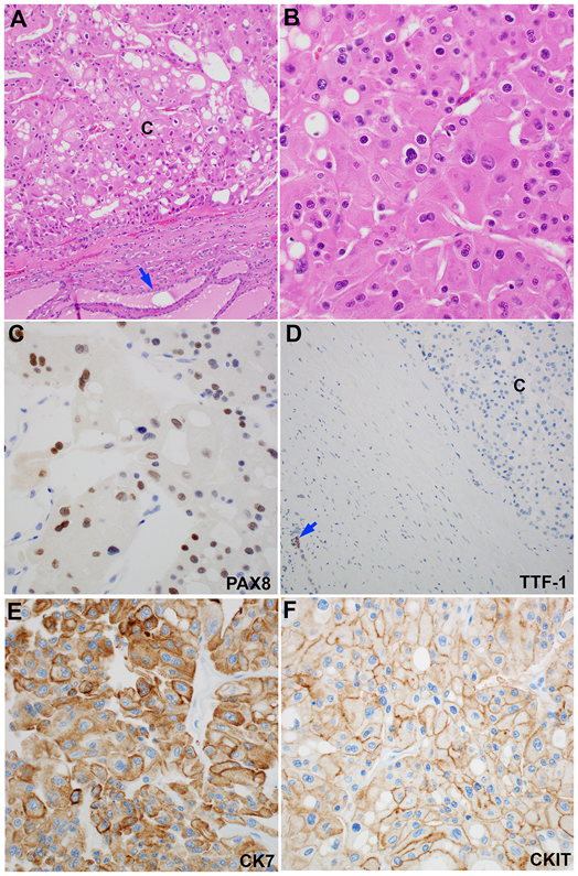
Figure 3. Metastatic chromophobe renal cell carcinoma to the thyroid gland.
(A) At low power (100X), the tumor is separated from the background thyroid (blue arrow) by a fibrous capsule. (B) At high power (400X), the tumor cells have wrinkled nuclei, binucleation, perinuclear halo, and abundant eosinophilic cytoplasm. (C-F) The tumor shows a PAX8-positive, TTF-1-negative, CK7-positive, and CKIT-negative immunoprofile. Note that the background thyroid is TTF-1 positive (blue arrow in panel D).

