. 2021 Jul 21;12(32):10901–10918. doi: 10.1039/d1sc03486c

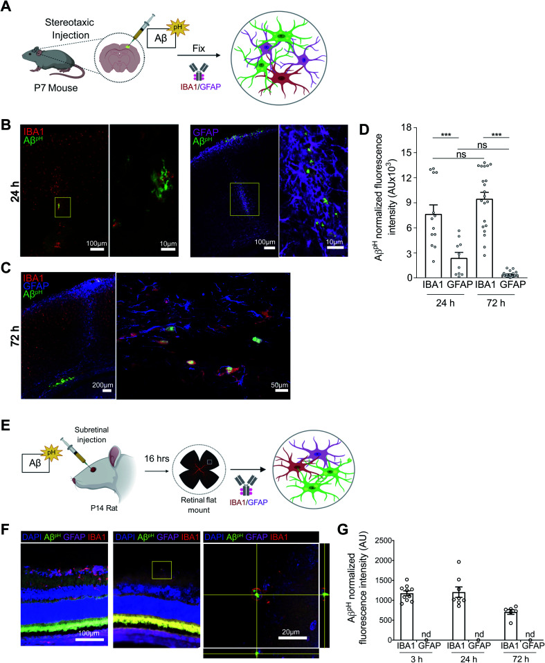
Fig. 4. AβpH is phagocytosed by cortical microglia and astrocytes in vivo and by rat retinal microglia in vivo. (A) Schematic of stereotaxic microinjection of AβpH in the somatosensory cortex of P7 mouse followed by staining of fixed tissue section after 24 and 72 hours. (B) Phagocytic uptake of AβpH by IBA1+ microglia and GFAP+ astrocytes in the periventricular white matter at the 24 hour timepoint. The box represents the region of the fluorescence image where high magnification confocal imaging was done. (C) IBA1+ microglia show bright green fluorescence at 72 hours in the same region indicating presence of AβpH within the cells at this timepoint. GFAP+ astrocytes do not show any green fluorescence in this region at this timepoint suggesting either degradation of the peptide or insufficient AβpH concentration for detectable phagocytic uptake by these cells. (D) Quantification of AβpH fluorescence within IBA1+ microglia and GFAP+ astrocytes located in the pia and white matter regions show more AβpH uptake by microglia compared to astrocytes. (E) Schematic of subretinal injection of AβpH to evaluate its in vivo uptake by rat retinal microglia and astrocytes. (F) IBA1+ rat retinal microglia phagocytose AβpHin vivo. (G) Quantification of AβpH uptake into retinal IBA1+ microglia and GFAP+ astrocytes. No fluorescence was detected in astrocytes at these 3 time points (n.d.). Data shown as mean ± s.e.m. from 2 animals.

Fig. 4