. 2021 Jul 21;12(32):10901–10918. doi: 10.1039/d1sc03486c

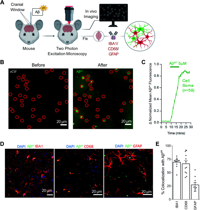
Fig. 5. AβpH is phagocytosed by microglia and astrocytes in vivo in the cerebral cortex. (A) Schematic of how AβpH phagocytic uptake is imaged through a cranial window in vivo in real time using two-photon excitation microscopy. (B) In vivo two-photon imaging of the mouse barrel cortex before and after topical application of AβpH. The fluorescence increases in cell somata (indicated by red circles) reflecting AβpH uptake. Shadows in the right image are created by the presence of pial and intraparenchymal vessels. (C) Quantification of mean AβpH fluorescence in cell somata over time. The data were normalized to the maximum mean AβpH fluorescence for each cell and then averaged. Data shown as mean ± s.e.m. N = 59 somata from 2 animals. (D) 1.5 to 3 hours after in vivo two-photon imaging of AβpH, animals were perfusion-fixed and cortical slices were stained for microglia, lysosomes/endosomes, and astrocytes using IBA1, CD68, and GFAP antibodies, respectively. (E) Quantification of AβpH colocalization with IBA1, CD68, and GFAP suggests that most AβpH is taken up by microglia and astrocytes in vivo. Data shown as mean ± s.e.m. N = 12 stacks from 3 animals.

Fig. 5