Figure 4.
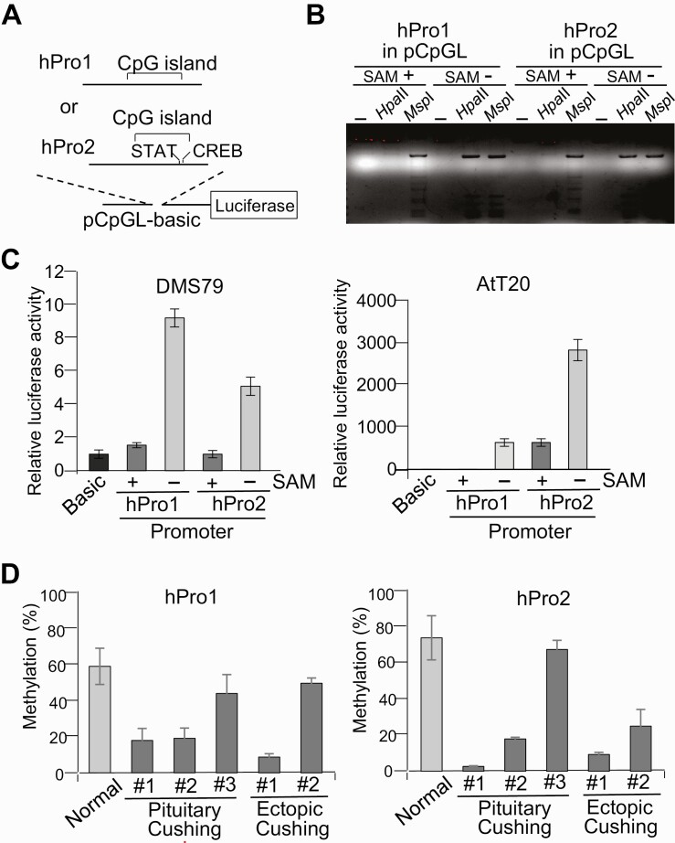
First and second proopiomelanocortin (POMC) promoter activity with DNA methylation. A, Structure of luciferase reporter plasmids using the dinucleotide 5′-CG-3′ (CpG) sequence free luciferase vector CpG-free luciferase reporter plasmid (pCpGL) and first (hPro1; –428 to +68) and second promoter (hPro2; +6657 to +7136) fragments. The relative position of the CpG island and potential STAT and CREB binding sites on these fragments are indicated. B, First (hPro1) and second (hPro2) promoter reporter plasmids treated with methyltransferase with (+) or without (–) S-adenosylmethionine (SAM), digested with no enzyme (–), Hpa II, or Msp I, and analyzed by agarose gel electrophoresis. C, Luciferase assays using first (hPro1) and second (hPro2) promoter reporter plasmids treated with (+) and without (–) SAM, with generated luciferase activities compared to negative control plasmid CpG-free luciferase reporter plasmid (pCpGLbasic) (Basic). D, Methylation levels in first (hPro1) and second (hPro2) promoters analyzed by bisulfite-conversion–based methylation-specific polymerase chain reaction using DNA isolated from 3 pituitary adrenocorticotropin (ACTH)-secreting tumors (pituitary #1, #2, and #3; see Fig. 1A) and 2 ectopic ACTH-secreting tumors (thymus #1 and lung #2; see Fig. 1A), with normal pituitary obtained at autopsy (normal) as control. Tumor characteristics are shown in Tables 1 and 2.

