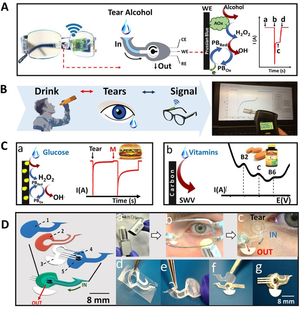
Figure 1. Eyeglasses-based Fluidic Device.

A) Photographic depiction and schematics of the fluidic device and wireless electronics integrated into the eyeglasses platform along with a representation of enzymatic alcohol detection and signal transduction, where, (a) corresponds to the baseline; (b) current change due the captured tear; (c) the measured alcohol signal and (d) drying of the device. B) Steps for tear alcohol detection consisting of ingestion of an alcoholic drink followed by tear stimulation and amperometric measurement. C) Representation of glucose (a) and vitamin detection (b). For the enzymatic glucose detection, the enzymatic reaction is presented showing the oxidation of tear glucose and hydrogen peroxide as byproduct along with its detection by the Prussian blue electrode. For the vitamin detection a typical SWV response is showed indicating the peak for vitamins B2, C and B6. D) Exploded view of fluidic device: (1) is the top polycarbonate membrane, (2) is the double adhesive spacer, (3) is the paper outlet, (4) the electrochemical (bio)sensor and (5) the bottom polycarbonate membrane. Tears stimulation: (a) Menthol tear stick (b) Volunteer applying the tear stick under the left eye. (c) Tear entering the inlet of the device. Fluidic device fabrication: (d) Adhesive spacer removed from PET substrate. (e) Spacer is placed on the bottom membrane. (f) Electrode and outlet are placed on top of spacer followed by top membrane. (g) Final device on eyeglasses nose-bridge pad.
