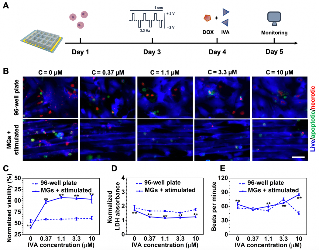
Figure 6.
Cardioprotective efficacy assessment of IVA in combination therapy with DOX. A) Schematic timeline of cardioprotective efficacy evaluations enabled by the heart-on-a-chip. B) Representative fluorescent images of CM viability treated by increasing dosages of IVA under the same DOX concentration (1.1 × 10⁻⁶ M). (Scale bar = 50 μm, CMs were seeded on Day 1, stimulated on Day 3, following the addition of DOX and IVA on Day 4, and imaged on Day 5.) C) CCK-8 assay for quantitative analysis of CM viability treated with increasing dosage of IVA under the same DOX concentration (1.1 × 10⁻⁶ M). D) LDH assay for quantitative analysis of CM viability treated by increasing dosage of IVA under the same DOX concentration (1.1 × 10⁻⁶ M). E) Quantification of beating behavior of CMs cultured under increasing dosages of IVA under the same DOX concentration (1.1 × 10⁻⁶ M) represented as beats per minute. (6C and 6D data are mean ± SEM, n ≥ 3; 6E data are mean ± SD, n = 3; *p < 0.05, **p < 0.01, ***p < 0.001).

