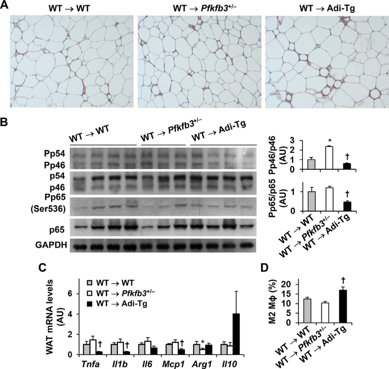
Fig. 3.
Adoptive transfer of wild-type hematopoietic cells to recipients does not weaken the effect of aP2-driven Pfkfb3 overexpression on ameliorating the severity of HFD-induced WAT inflammation. (A) WAT macrophage infiltration. (B) WAT proinflammatory signaling. (C) WAT expression of genes for cytokines and inflammatory mediators. (D) WAT macrophage alternative (M2) activation. For A–D, chimeric mice were generated as described in Fig. 1 and fed an HFD for 12 wk. For A, WAT sections were stained for F4/80 expression. For B, WAT lysates were subjected to Western blot analysis. Left panels, representative blots from 3–4 WAT per group. Bar graphs, quantification of blots for all WAT samples. n=7. For C, WAT RNA was subjected to reverse transcription and real-time PCR. n=7. For D, WAT macrophage alternative activation was examined using FACS analysis. Stromal cells isolated from WAT in chimeric mice were analyzed for mature macrophages (F480+ CD11b+ cells), which were further analyzed for M2 macrophages (CD206+ CD11c− cells). n=5–7. For bar graphs in B,C, and D, data are means ± SEM. *, P < .05 WT → Pfkfb3+/− vs. WT → WT for the same protein (B) or gene (C); †, P < .05 WT → Adi-Tg vs. WT → WT for the same protein (B), gene (C), or macrophage type (D).

