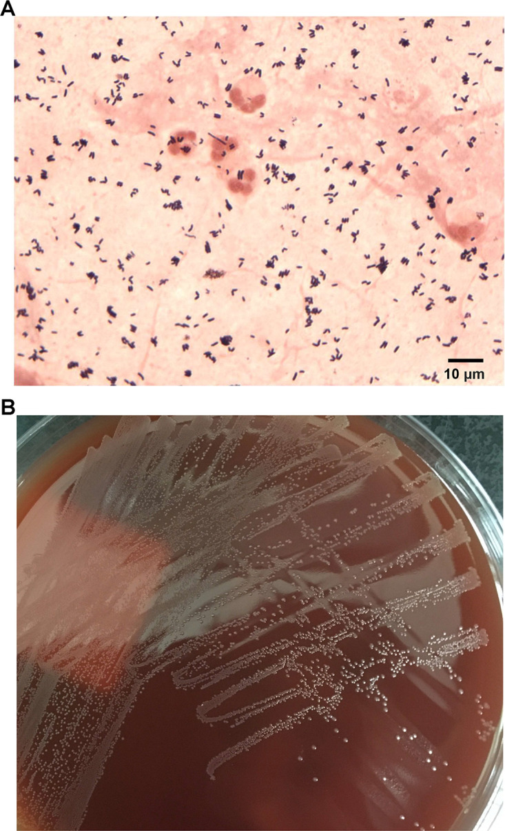
FIG 2.

Microscopic and macroscopic morphology of Corynebacterium accolens. (A) Direct Gram stain of tracheal aspirate demonstrating coryneform Gram-positive rods and polymorphonuclear leukocytes. (B) A subculture of the C. accolens isolate, showing small, gray, nonhemolytic colonies on Columbia sheep blood agar after 48 h of incubation at 35°C with 5% CO2.
