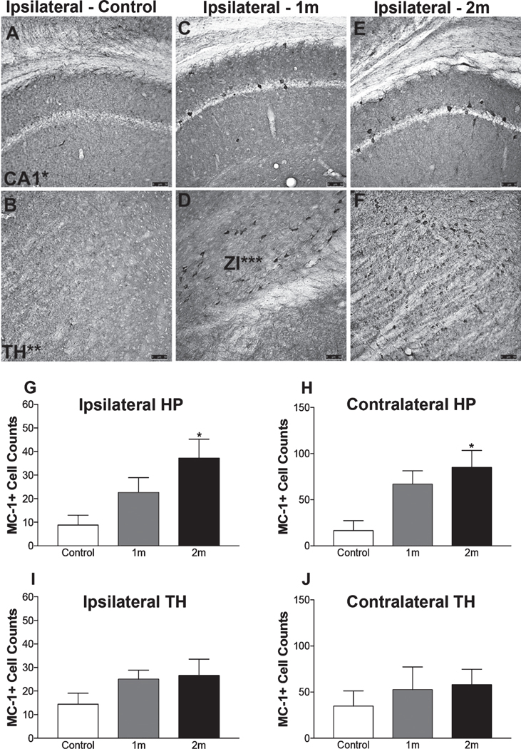
Fig. 5.
Deposits of pathogenically misfolded tau are present in the brain 2m post-injection. Representative photomicrograph of a MC-1 stained mouse section, no MC-1 immunoreactivity was observed in the brains of control mice (n=5/timepoint; scale bar, 100 μm). Representative photomicrographs of MC-1 immunoreactivity in the ipsilateral hippocampus and ipsilateral TH obtained from a control (A-B), 1m (C-D), and 2m (E-F) NiPSCE injected mice (n=5/timepoint; scale bar, 100 μm). Quantification of MC-1+cells (G-J) in NiPSCE injected mouse brains and controls shows that MC-1 immunoreactivity was significantly increased in the ipsilateral hippocampus (G), and ipsilateral TH (I) of mice sacrificed at 2m post-injection as compared to control mice. (n=5/group/timepoint; One-way ANOVA with Bonferonni post hoc test, *p<0.05 versus control).

