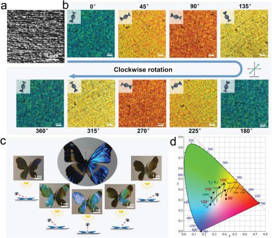
Figure 2.

Optical properties of the CNCs@glucose layer. a) SEM cross‐section image of the film showing evident cholesteric structure of CNCs. b) POM‐λ images of the film showing a periodic color change from blue to orange following the clockwise rotation. c) The angle‐related structural colors of an artificial “Morpho Helena”. d) CIE 1931 color space chromaticity diagrams for the LC layer during the color‐changing process, coordinate values of which were obtained for every 5° increment in the view angle.
