Figure 3.

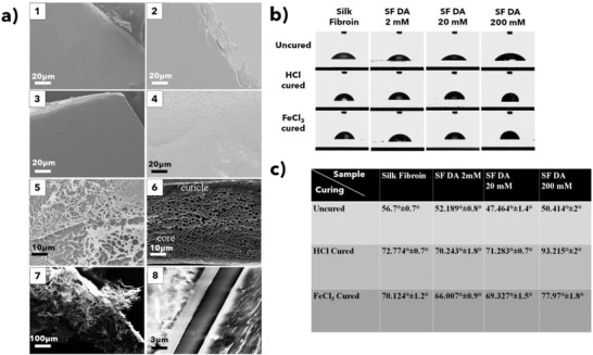
a1–a4) SEM images and relative film insets of different SF–PDA blends. a1) SF film; a2) SF–PDA 2 × 10⁻³ m; a3) SF–PDA 20 × 10−3 m; a4) SF–PDA 200 × 10−3 m; a5) alveolar porous structure of SF–PDA 200 × 10−3 m after curing with FeCl3 30 × 10−3 m and lap‐shear test; a6) natural byssal plaque of Mytilus edulis; a7) alveolar structure of SF–PDA 200 × 10−3 m cured with FeCl3 30 × 10−3 m (not subjected to lap‐shear test) with a lower magnification; a8) cross‐sectional SEM of SF–PDA 200 × 10−3 m between two glass slides. Markers are reported for each image. a6) Reproduced with permission.[ 1 ] Copyright 2005, Taylor & Francis. b,c) Static contact angles of SF and SF–PDA films uncured or cured either with 4 µL of HCl 55 × 10−3 m or FeCl3 30 × 10−3 m against bidistilled water.
