Image.

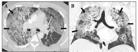
Computed tomography angiography of the chest. Black arrows indicate ground glass opacities. (A) Axial image showing bilateral central and peripheral ground-glass opacities. (B) Coronal image demonstrating bilateral ground-glass opacities in upper and lower lung fields.
