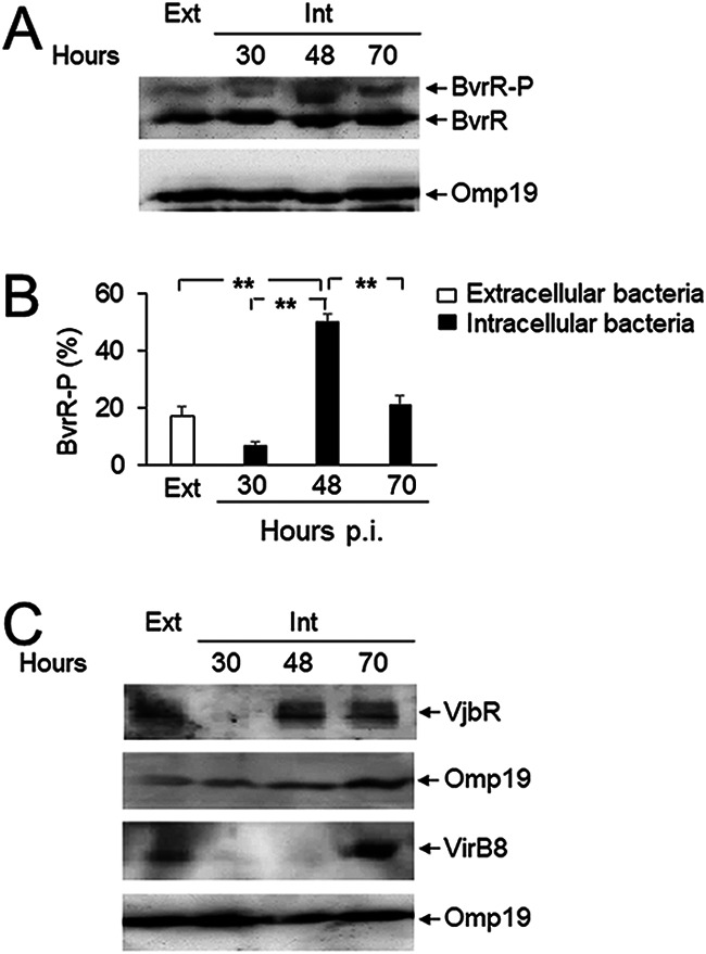
FIG 5.

Increased activation of BvrR/BvrS and expression of vjbR and virB at late stages of the intracellular infection cycle. (A) RAW 264.7 macrophages were infected with B. abortus, and at the indicated times, intracellular bacteria were extracted and purified (Int). Bacterial lysates were prepared and separated by 10% Phos-tag SDS-PAGE, transferred to PVDF membranes, and probed with anti-BvrR or anti-Omp19 (loading control) antibodies. Bacteria grown in TSB in vitro were used as a control (Ext). (B) The percentage of total BvrR that was phosphorylated (BvrR-P) was calculated for each indicated condition by densitometry from at least three independent experiments. Statistical significance was calculated by analysis of variance and Tukey’s multiple-comparison test (**, P < 0.005). (C) Lysates from intracellular bacteria obtained as described for panel A were used for the detection of VjbR and VirB8 by Western blotting. These results are representative of three independent experiments.
