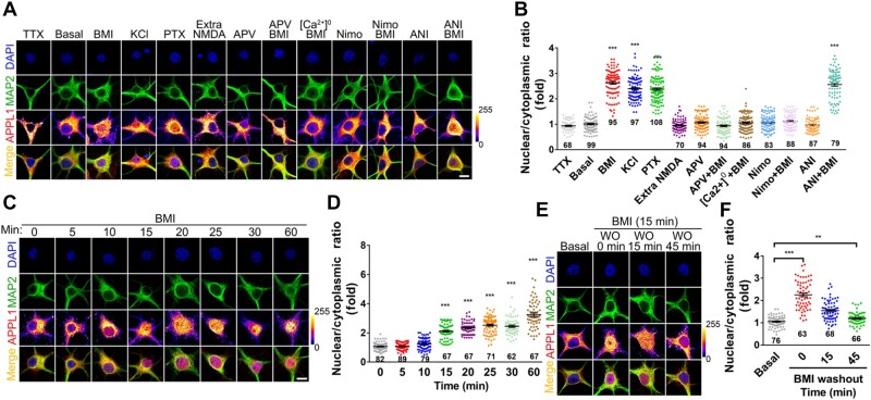
Figure 1.
Synaptic activity induces nuclear accumulation of APPL1 in the cultured hippocampal neurons. (A) Cultured hippocampal neurons at DIV 14‒17 were untreated (basal) or treated with Tetrodotoxin (TTX, 1 μM), BMI/4-AP (BMI, 50 μM/2.5 mM), Picrotoxin (PTX, 50 μM), KCl (50 mM), extrasynaptic NMDAR activation protocol (Extra NMDA), APV (50 μM), Nimodipine (Nimo, 10 μM), or Anisomycin (ANI, 50 μM), respectively, for 1 h. Alternatively, hippocampal neurons were pretreated with APV, Nimodipine, calcium-free extracellular media ([Ca2+]0), or Anisomycin, respectively, for 30 min followed by treatment with BMI for another 1 h. After treatment, the cultures were immunostained with antibodies against MAP2 (green) and APPL1 (color lookup table, pixel intensities from 0 to 255) and with DAPI nuclear dye (blue). Scale bar, 10 μm. (B) Statistical analysis of the nuclear/cytoplasmic ratio of APPL1. (C) Hippocampal neurons at DIV 14‒17 were pretreated with Leptomycin B (10 nM) for 1 h, then treated with BMI for indicated times, and subsequently stained with antibodies against MAP2 (green) and APPL1 (color lookup table) and with DAPI nuclear dye (blue). Scale bar, 10 μm. (D) Statistical analysis of the nuclear/cytoplasmic ratio of APPL1. (E) Hippocampal neurons were treated with BMI for 15 min, washed out (WO), and then incubated in fresh media absent of BMI for different times as indicated. Neurons were stained with antibodies against MAP2 (green) and APPL1 (color lookup table) and with DAPI nuclear dye (blue). Scale bar, 10 μm. (F) Statistical analysis of the nuclear/cytoplasmic ratio. **P < 0.01, ***P < 0.005.

