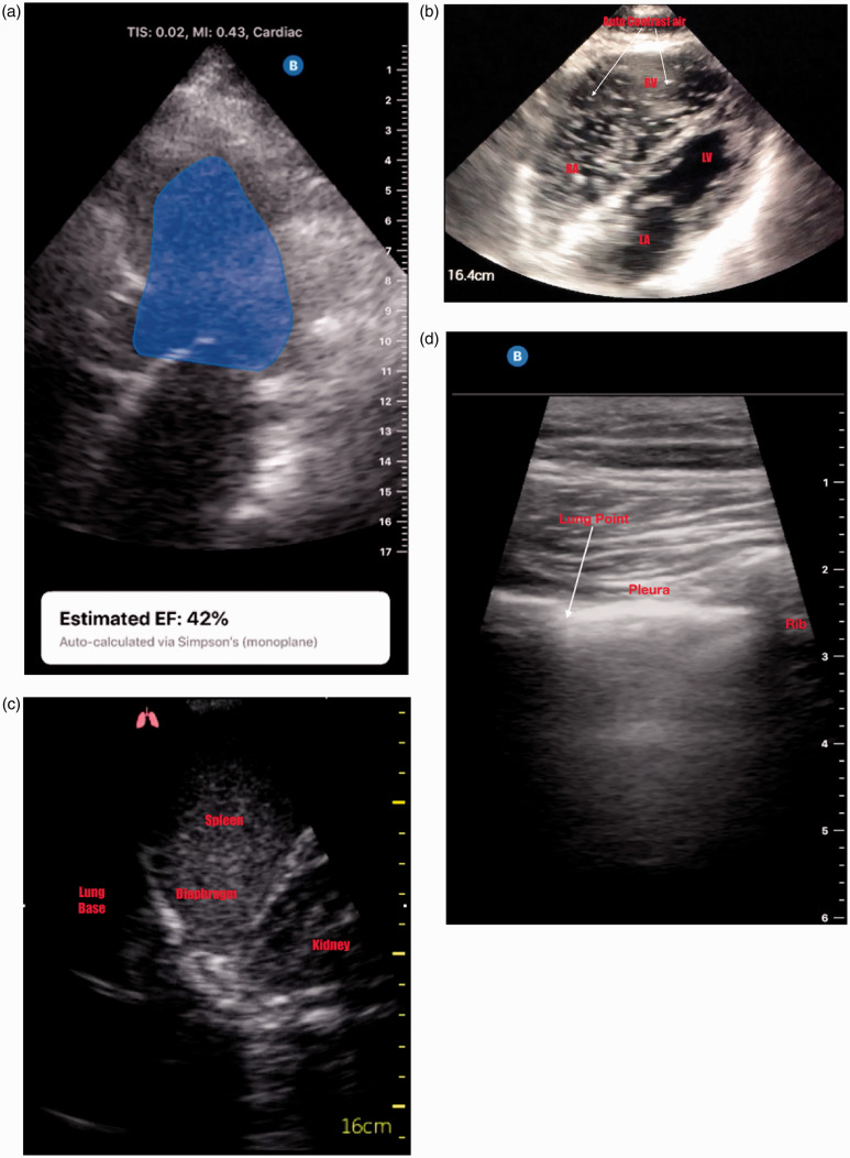
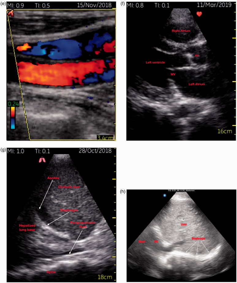
Figure 2.
Various images obtained from two popular portable handheld devices. (a) Apical four-chamber view with auto ejection fraction calculation. (b) Apical four-chamber view with saline auto-contrast demonstration in right heart during adrenaline minijet administration for cardiac arrest. (c) Left lung base scan demonstrating relevant anatomy. (d) Scan of the upper chest wall demonstrating a lung point where normal pleural sliding meets the interface with the non-sliding part of the pneumothorax. (e) Colour flow Doppler across jugular vein and carotid artery. (f) Normal parasternal long axis view of the heart. (g) Right lung base/right upper quadrant view showing cirrhotic liver, ascites and parapneumonic fluid. (h) Subcostal diaphragm scan used later to perform M-mode for excursion fraction calculation in a weaning patient.


