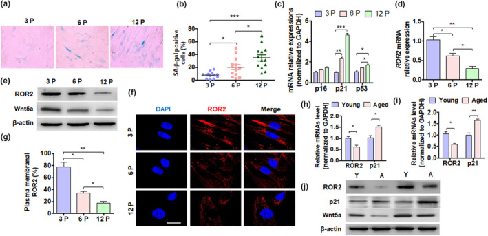
FIGURE 1.

ROR2 expression is reduced in aged DPSCs. (a) DPSC aging was induced by cell passage. SA β‐gal staining of DPSCs from young donors at passage 3 (3P), 6 (6P), and 12 (12P). (b) Quantitative analysis of SA β‐gal‐positive DPSCs (3P, 6P, and 12P). (c and d) RT‐qPCR was performed to detect the p16, p21, p53, and ROR2 mRNA expression in DPSCs (3P, 6P, and 12P). (e) Western blot analysis was performed to detect the ROR2 protein level in DPSCs from young donors. (f) Immunofluorescence staining for ROR2 in DPSCs; scale bar: 20 µm. (g) Quantitative analysis of cells with ROR2 in the plasma membrane of (f). (h and i) RT‐qPCR was performed to detect the ROR2 and p21 mRNA expression in the dental pulp tissues of young and elderly patients. (h) DPSC isolation (6P) from young and aged donors. (j) Western blot analysis of ROR2 and p21 expression in DPSCs (6P) isolated from young (Y) and aged (A) donors. For all analyses, *p < 0.05, **p < 0.01, and ***p < 0.001 compared to their corresponding controls
