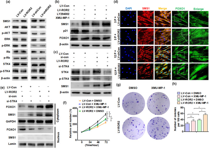
FIGURE 6.

STK4‐FOXO1 axis mediates ROR2‐regulated SMS1 expression in DPSCs. (a) DPSCs at 3P were transfected with LV‐shCon and LV‐shROR2, while at 12P were transfected with LV‐Con and LV‐ROR2; Western blot analysis was performed to detect the indicated protein expression. (b) DPSCs (12P) were transfected with LV‐ROR2 or LV‐Con, and treated with AKT pathway inhibitor (LY294002) or STK4 inhibitor (XMU‐MP‐1) for 2 h; Western blot analysis was performed to detect SMS1, p21, and FOXO1 protein expression. (c) DPSCs (12P) were transfected with LV‐ROR2, si‐STK4, or their control vectors, and RNA; Western blot analysis was performed to detect the indicated protein level. (d) Immunofluorescence staining for SMS1 and FOXO1 in DPSCs at 3P and 12P after being transfected with LV‐shROR2 or their control vector; scale bar: 100 µm. (e) Western blot analysis was performed to detect FOXO1 and SMS1 in subcellular fractionations of DPSCs (12P) transfected with LV‐ROR2, si‐STK4, or their control vector, and RNA; Lamin and GAPDH served as controls for the nuclear and cytoplasmic fractions, respectively. (f) DPSCs at 12P were treated with XMU‐MP‐1 after LV‐ROR2 transfection for 48 h, and an MTS assay was performed to detect cell proliferation. (g and h) CFU assay was performed in DPSCs at 12P after transfection and treatment, and crystal violet was used to stain the colonies on day 14. DPSCs at passages 3–12 used in these experiments were from young donors. For all analyses, *p < 0.05, **p < 0.01, and ***p < 0.001 compared to their corresponding controls
