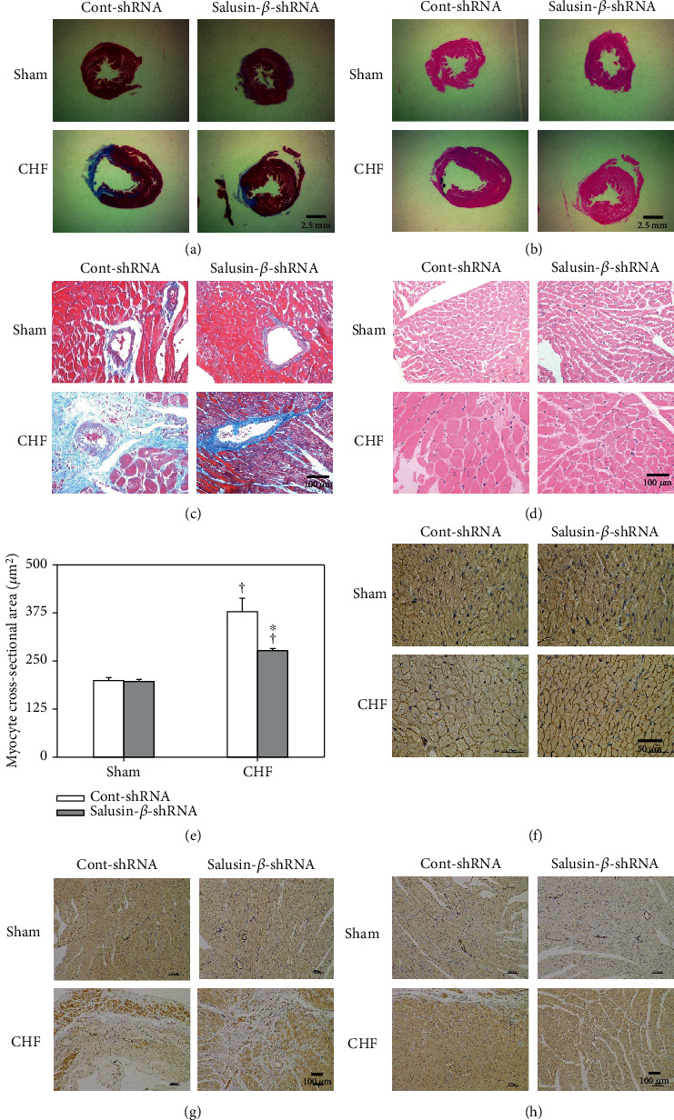
Figure 6.

Effects of salusin-β knockdown on left ventricular remodeling and microvascular density. Sections of myocardium with Masson's stain under low- (a) and high-power microscope (c) showing fibrosis. Sections of myocardium with HE staining under low- (b) and high-power microscopy (d) and dystrophin staining (f) showing the size of cardiomyocytes. Bar graph showing the quantitative analysis of the cross-sectional area of cardiomyocytes (e). Sections of myocardial infarct border (g) and remote zone (apex of heart) (h) with endothelial marker CD31 immunohistochemistry staining showing the microvascular density. Values are mean ± SE. ∗P < 0.05 compared with Cont-shRNA, †P < 0.05 compared with Sham. n = 6 for each group.
