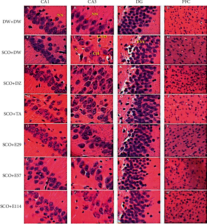
Figure 6.

Photographs of the hippocampal and prefrontal cortex sections after hematoxylin and eosin staining (250X). Hippocampus (CA1, CA3, and DG); PFC: prefrontal cortex; CA1 and 3: Cornu ammonis 1 et 3; DG: dentate gyrus; N: neuron; NN: normal neuron; GVD: granulovacuolar degeneration; S spongiosis; CH: chromatolysis; PVE: perivascular edema; DW: distilled water (10 ml/kg); SCO: scopolamine (1 mg/kg); DZ: donepezil (1.2 mg/kg); TA: tacrine (10 mg/kg); and E29, E57, and E114: aqueous extract of Z. jujuba at respective doses of 29, 57, and 114 mg/kg. DW + DW: normal control group; SCO + DW: negative control group; SCO + DZ: positive control group treated with donepezil; SCO + TA: positive control group treated with tacrine; and SCO + E29-E114: test groups treated with the extract of Z. jujuba.
