Figure 2.

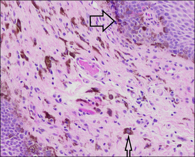
Biopsy, distal esophagus: basal layer proliferation of melanocytes, noted by their prominent nucleoli and adjacent melanin pigment (large arrow). Submucosal melanophages are visible, with notable pigment inclusion (small arrow).

Biopsy, distal esophagus: basal layer proliferation of melanocytes, noted by their prominent nucleoli and adjacent melanin pigment (large arrow). Submucosal melanophages are visible, with notable pigment inclusion (small arrow).