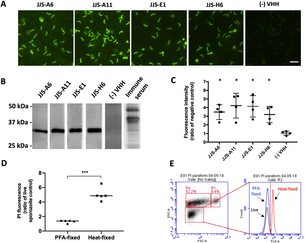
Fig. 2.
Discovery of a ~34 kDa protein target exposed on the surface of Cryptosporidium parvum sporozoites bound by single-domain camelid antibodies (VHHs). (A) Fluorescent micrographs (600x) localizing FITC-labeled VHH monomers bound to permeabilized sporozoites. Scale bar = 5 μm. (B) An ~34 kDa protein band of separated C. parvum lysate bound by VHH monomers. The experiment was performed in the presence of a non-specific VHH as a negative control and immune alpaca serum as a positive control. (C) Detection of VHH monomers binding the surface of non-permeabilized sporozoites by flow cytometry corroborates findings from microscopy (Mann-Whitney test; *P=0.02). Data is adjusted to ‘no VHH’ control. Values indicate means and error bars indicate S.D. (n=4). (D) Sporozoites fixed with paraformaldehyde remain impermeable to propidium iodide (PI) in contrast to sporozoites fixed with heat as measured by flow cytometry (t-test, P<0.0001). Data points indicate the ratio of the PI signal generated by live sporozoites (n=5). PFA, paraformaldehyde.(E) A single replicate representative of flow cytometry output indicating the intensity of the PI fluorescence signal detected in FLA-3 channel for live, PFA-fixed and heat-permeabilized sporozoites. Sporozoites are pre-gated in excysted oocyst preparation, where R1 represents full oocysts, R2 sporozoites and R4 oocysts in different stages of excystation.

