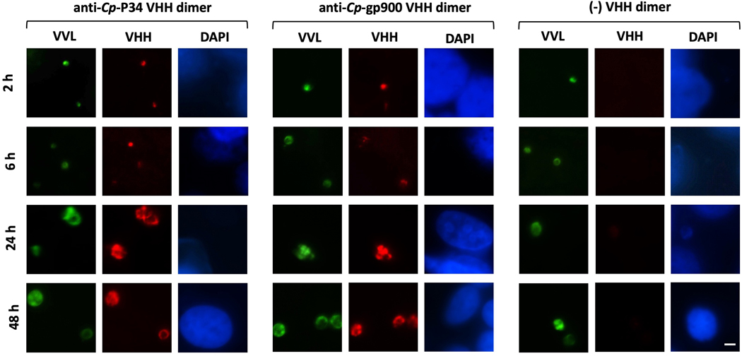
Fig. 5.
Expression pattern of P34 (Cp-P34) and gp900 (Cp-gp900) of Cryptosporidium parvum during intracellular development in vitro. Fluorescent micrographs (600x) demonstrating Madin-Darby bovine kidney (MDBK) cells with intracellular stages of C. parvum over the course of infection- at 2, 6, 24 and 48 h p.i. Parasites are stained green with FITC-labeled Vicia villosa lectin (VVL); binding of the anti-Cp-P34 VHH dimer (JJS-A6/JJS-A11) and anti-Cp-gp900 VHH dimer (JMP-F7/JMP-F7) are detected in red with TRITC-labeled secondary antibody and the cell nucleus is counter-stained in blue with DAPI. A non-specific VHH dimer is included as a negative control. Scale bar = 5 μm.

