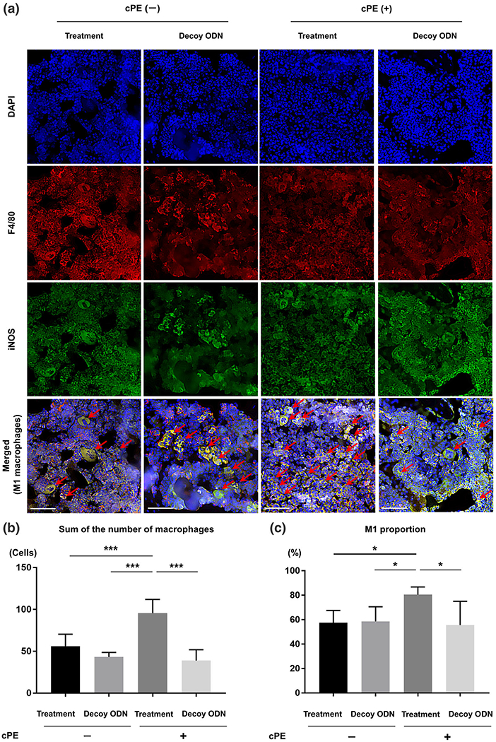
FIGURE 4.

Photomicrographs and graphs showing M1 pro-inflammatory macrophages. Immunofluorescence staining of anti-F4/80 antibody (red) and anti-inducible nitric oxide synthase (iNOS) antibody (green) was used to identify the M1 pro-inflammatory macrophages. DAPI (blue) was used to visualize nuclei. In merged images, the cells double positively stained with both anti-F4/80 antibody and anti-iNOS antibody indicated M1 pro-inflammatory macrophages (arrow) (a). In this section, quantitative assessments of macrophages (the sum of the number of cells positively stained with F4/80 and the number of cells double positively stained with both F4/80 and iNOS) (b) and the proportion of M1 pro-inflammatory macrophages (based on the following calculation: the number of cells double positively stained with both F4/80 (red) and iNOS divided by the sum of the number of cells positively stained with F4/80 and the number of cells double positively stained with both F4/80 and iNOS) (c) were also shown based on manually counting in three randomly selected views of each specimen with ×200 magnification by ImageJ software. White bar: 50 μm. *p < 0.05, **p < 0.001, ***p < 0.0001
