
Figure 4. NAD+ redox imbalance exacerbates diabetic cardiomyopathy, independent on tissue fibrosis.
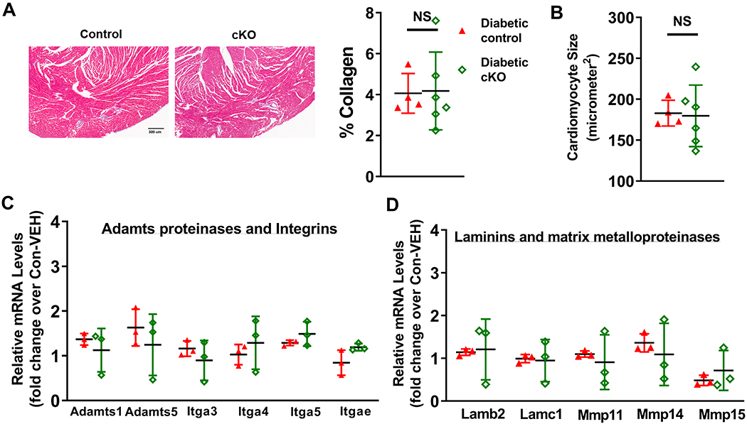
(A) Collagen levels of diabetic mouse hearts were quantified by trichrome staining. (B) Cardiomyocyte sizes of these hearts were quantified. N=4-6. Transcript expression levels of fibrosis-related genes were quantified by qPCR analyses. (C) Adamts proteinases and integrins, and (D) laminins and MMPs in diabetic control and diabetic cKO male hearts were measured. N=3.