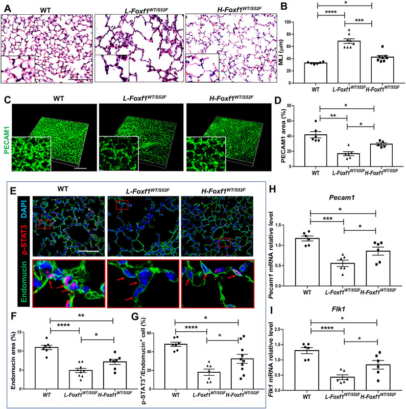
Figure 4. Low body weight in juvenile Foxf1WT/S52F mice is associated with alveolar capillary dysplasia and decreased p-STAT3.
(A-B) H&E staining of lung sections shows severe alveolar simplification in L-Foxf1WT/S52F mice. Mean linear intercept (MLI) was determined using 5 random images from each of 3 lung sections per mouse (n=6-8 mice per group). Scale bars are 50μm. (C-D) Whole mount PECAM1 immunostaining of lung slices shows decreased capillary density in L-Foxf1WT/S52F lungs. Data were quantified using 5 random lung images per mouse (n=5-6 mice per group). Scale bars are 100μm. (E-G) Immunostaining for Endomucin (green) and p-STAT3 (red) shows decreased capillary density and a decreased number of endothelial cells expressing p-STAT3 in L-Foxf1WT/S52F lungs. Bottom images show the area in red box. Arrows point to nuclei of endothelial cells. Slides were counterstained with DAPI (blue). Five random lung images per mouse were used to quantify the data (n=7-10 mice per group). Scale bars are 100μm. (H-I) qRT-PCR shows that Pecam1 and Flk1 mRNAs are decreased in total lung RNA from L-Foxf1WT/S52F mice. mRNAs were normalized using β-actin mRNA (n=6 mice per group). NS indicates no significance, * indicates p < 0.05, ** indicates p < 0.01, *** indicates p < 0.001, **** indicates p < 0.0001.

