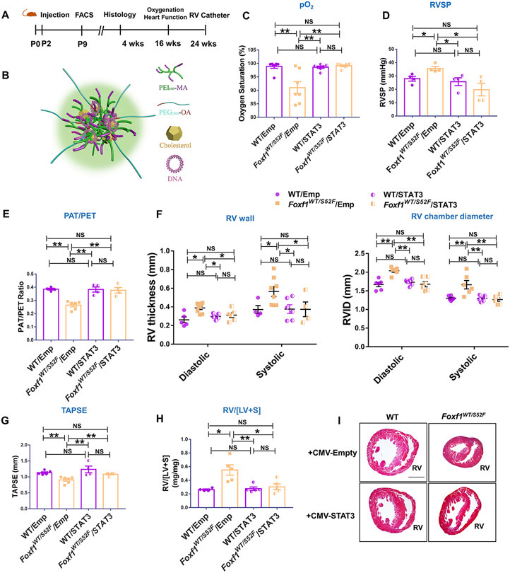
Figure 7. Nanoparticle delivery of Stat3 cDNA prevents RV hypertrophy and increases survival of Foxf1WT/S52F mice.
(A) Schematic representation of nanoparticle treatment of Foxf1WT/S52F mice. Nanoparticle containing either CMV-Stat3 (STAT3) or a CMV empty plasmid (Emp) were injected at P2 via the facial vein. FACS analysis was performed at P9. Histology and immunostaining were carried out at P28. Echocardiography and measurements of arterial oxygenation were performed at 4 months of age, whereas RV catheterization was performed at 6 months of age. (B) Schematic representation of nanoparticle structure shows PEI and PEG polymers, cholesterol and plasmid DNA. (C) Measurements of arterial oxygenation show that nanoparticle delivery of Stat3 DNA increases pO2 in Foxf1WT/S52F mice (n=7 mice per group). (D) STAT3 treatment decreases RVSP in Foxf1WT/S52F mice (n=4 mice per group). (E-G) Echocardiography shows improved ratio of pulmonary acceleration time (PAT) to pulmonary ejection time (PAT/PET) in Foxf1WT/S52F mice injected with STAT3 nanoparticles compared to CMV empty controls. Nanoparticle delivery of Stat3 cDNA increases TAPSE but decreases diastolic and systolic RV wall thickness and the right ventricular internal diameter (RVID) in Foxf1WT/S52F mice (n=4-8 mice per group). (H) Nanoparticle delivery of Stat3 cDNA decreases RV/[LV+S] in P28 Foxf1WT/S52F mice (n=4-5 mice per group). (I) H&E staining shows transverse heart sections of WT and Foxf1WT/S52F mice that were either treated with STAT3 or control nanoparticles. Scale bars are 0.8mm. NS indicates no significance, * indicates p < 0.05, ** indicates p < 0.01. Abbreviations: RV/[LV+S], ratio of right ventricle weight to the combined weight of left ventricle and interventricular septum; RV, right ventricle; TAPSE, Tricuspid annular plane systolic excursion.

