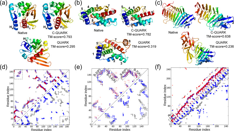
Fig. 3. Illustrative examples for contact-guided ab initio structure folding.
a–c 3D structures for 2d7jA [10.2210/pdb2D7J/pdb], 1y9iA [10.2210/pdb1Y9I/pdb] and 4peuA [10.2210/pdb4PEU/pdb], with blue to red running from N- to C-terminal. d–f corresponding contact-maps for the native structures (gray circles), predicted contacts (red circles), the contacts in the C-QUARK models (blue circles in the upper left triangles), and contacts in the QUARK models (blue circles in the lower right triangle). In a, d, HN and HC represent the N- and C-terminal helices, respectively, which are in contact in the native structure and C-QUARK model but not in the QUARK model, as highlighted in rectangles in the contact-map. In b, e, numbers indicate the order of helices from N- to C-terminal, where the contacts that contribute to the correct helix packing are highlighted by rectangles with ‘A’ and ‘P’ referring to anti-parallel and parallel, respectively, in the contact-map. In c, f, numbers indicate the order of the beta-strands at the N-terminal whose contacts are highlighted by rectangles in the contact-map.

