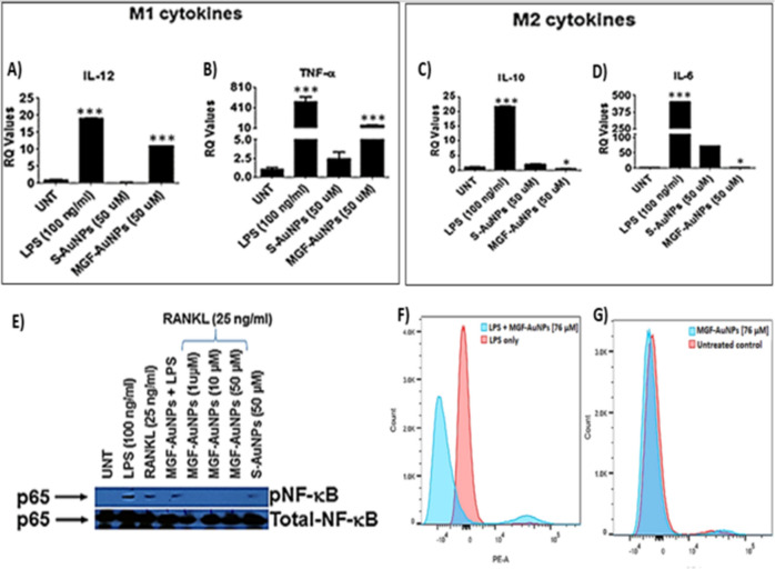
Figure 8.
MGF-AuNPs induced polarization of macrophages and inhibits NF-κB activation. (A–D) RAW 264.7 cells were pretreated with either Starch-AuNPs (S-AuNPs as control), or MGF-AuNPs for 2 h and treated either with LPS (100 ng/mL) or RANKL (25 ng/mL) or left untreated for 4 h. RNA was isolated from treated and untreated samples and analyzed for IL-12, TNF-α, IL-10, and IL-6 by real time PCR using probes from TaqMan, Applied Biosystems. (E). RAW 264.7 cells were either treated with LPS (100 ng/mL) or Starch-AuNPs (S-AuNPs as control), or MGF-AuNPs or left untreated for 30 min. The cells were lysed with 1X Lamellae buffer and lysates were run on PAGE gel and transferred onto nitrocellulose membranes. The membranes were than probed for either phospho- NF-κB or NF-κB using respective antibodies. (F). The RAW 264.7 cells were cultured overnight in 6 well plates and pre-treated with different doses of MGF-AuNPs (0, 32 µg/mL) for 3 h. Subsequently the cells were washed and treated with LPS (100 ng/mL) for 45 min. After incubation with LPS the cells were washed, fixed and permeabilized. After permeabilization the cells were stained with PE conjugated anti-NF-κB antibody for 45 min. The cells were washed and analyzed using flow cytometry. (G) The RAW 264.7 cells were cultured overnight in 6 well plates and pre-treated with different doses of MGF-AuNPs (0, 32 µg/mL) for 45 min. Cells were fixed, permeabilized and stained with PE conjugated anti-NF-κB antibody for 1 h and analyzed by flow cytometry.

