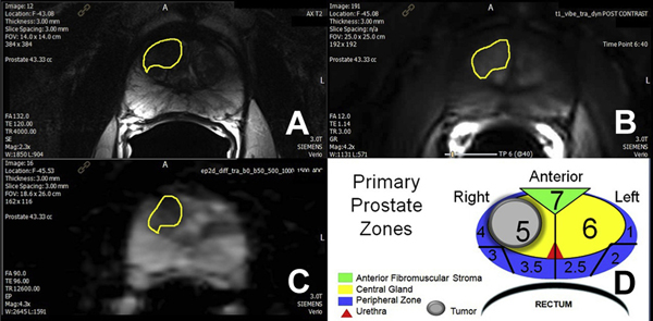
Figure 1.
Prostate MP-MRI in 66-year-old male with PSA 5.6 ng/ml. A, T2-weighted axial image shows anterior right central lesion with charcoal sign (yellow outline). B, dynamic contrast enhanced with type 3 focal enhancement curve. C, ADC map with ADC value 487 × 10−6 × mm2 per second. D, primary prostate zones with MR tumor volume (1.5 × 1.4 × 1.3 cm) = 1.4 cm3 and target core calculated volume (11 mm cancer) = 0.7 cm3.

