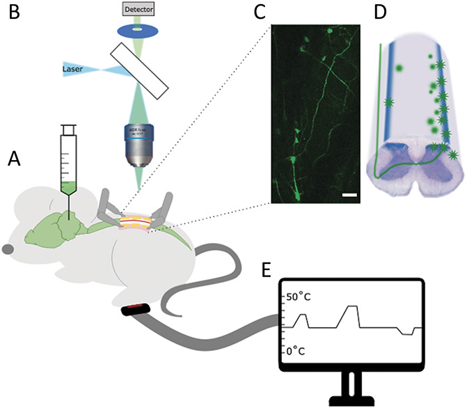
Figure 1.

Description of experimental setup. (A) Mice were injected with AAV9 expressing GCaMP at least 5 days before imaging. (B) Mice were imaged with a one-photon microscope and (C) labelled cells visualised in the dorsal horn of the spinal cord. Scale bar = 100 μm. (D) Cartoon of cellular labelling after parabrachial injections. During imaging sessions the ipsilateral peripheral paw of the mouse was stimulated electrically (with a cuff electrode around the sciatic nerve), mechanically (through brush or pinch), or (E) thermally (using a Peltier device applied to the plantar surface of the paw). AAV, adeno-associated virus.
